In biochemistry, flavin adenine dinucleotide (FAD) is a redox-active coenzyme associated with various proteins, which is involved with several enzymatic reactions in metabolism. A flavoprotein is a protein that contains a flavin group, which may be in the form of FAD or flavin mononucleotide (FMN). Many flavoproteins are known: components of the succinate dehydrogenase complex, α-ketoglutarate dehydrogenase, and a component of the pyruvate dehydrogenase complex.
FAD can exist in four redox states, which are the flavin-N(5)-oxide, quinone, semiquinone, and hydroquinone.[1] FAD is converted between these states by accepting or donating electrons. FAD, in its fully oxidized form, or quinone form, accepts two electrons and two protons to become FADH2 (hydroquinone form). The semiquinone (FADH·) can be formed by either reduction of FAD or oxidation of FADH2 by accepting or donating one electron and one proton, respectively. Some proteins, however, generate and maintain a superoxidized form of the flavin cofactor, the flavin-N(5)-oxide.[2][3]
Flavoproteins were first discovered in 1879 by separating components of cow's milk. They were initially called lactochrome due to their milky origin and yellow pigment.[4] It took 50 years for the scientific community to make any substantial progress in identifying the molecules responsible for the yellow pigment. The 1930s launched the field of coenzyme research with the publication of many flavin and nicotinamide derivative structures and their obligate roles in redox catalysis. German scientists Otto Warburg and Walter Christian discovered a yeast derived yellow protein required for cellular respiration in 1932. Their colleague Hugo Theorell separated this yellow enzyme into apoenzyme and yellow pigment, and showed that neither the enzyme nor the pigment was capable of oxidizing NADH on their own, but mixing them together would restore activity. Theorell confirmed the pigment to be riboflavin's phosphate ester, flavin mononucleotide (FMN) in 1937, which was the first direct evidence for enzyme cofactors.[5] Warburg and Christian then found FAD to be a cofactor of D-amino acid oxidase through similar experiments in 1938.[6] Warburg's work with linking nicotinamide to hydride transfers and the discovery of flavins paved the way for many scientists in the 40s and 50s to discover copious amounts of redox biochemistry and link them together in pathways such as the citric acid cycle and ATP synthesis.
Flavin adenine dinucleotide consists of two portions: the adenine nucleotide (adenosine monophosphate) and the flavin mononucleotide (FMN) bridged together through their phosphate groups. Adenine is bound to a cyclic ribose at the 1' carbon, while phosphate is bound to the ribose at the 5' carbon to form the adenine nucledotide. Riboflavin is formed by a carbon-nitrogen (C-N) bond between the isoalloxazine and the ribitol. The phosphate group is then bound to the terminal ribose carbon, forming a FMN. Because the bond between the isoalloxazine and the ribitol is not considered to be a glycosidic bond, the flavin mononucleotide is not truly a nucleotide.[7] This makes the dinucleotide name misleading; however, the flavin mononucleotide group is still very close to a nucleotide in its structure and chemical properties.


FAD can be reduced to FADH2 through the addition of 2 H+ and 2 e−. FADH2 can also be oxidized by the loss of 1 H+ and 1 e− to form FADH. The FAD form can be recreated through the further loss of 1 H+ and 1 e−. FAD formation can also occur through the reduction and dehydration of flavin-N(5)-oxide.[8] Based on the oxidation state, flavins take specific colors when in aqueous solution. flavin-N(5)-oxide (superoxidized) is yellow-orange, FAD (fully oxidized) is yellow, FADH (half reduced) is either blue or red based on the pH, and the fully reduced form is colorless.[9][10] Changing the form can have a large impact on other chemical properties. For example, FAD, the fully oxidized form is subject to nucleophilic attack, the fully reduced form, FADH2 has high polarizability, while the half reduced form is unstable in aqueous solution.[11] FAD is an aromatic ring system, whereas FADH2 is not.[12] This means that FADH2 is significantly higher in energy, without the stabilization through resonance that the aromatic structure provides. FADH2 is an energy-carrying molecule, because, once oxidized it regains aromaticity and releases the energy represented by this stabilization.
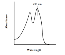
The spectroscopic properties of FAD and its variants allows for reaction monitoring by use of UV-VIS absorption and fluorescence spectroscopies. Each form of FAD has distinct absorbance spectra, making for easy observation of changes in oxidation state.[11] A major local absorbance maximum for FAD is observed at 450 nm, with an extinction coefficient of 11,300 M−1 cm−1.[13] Flavins in general have fluorescent activity when unbound (proteins bound to flavin nucleic acid derivatives are called flavoproteins). This property can be utilized when examining protein binding, observing loss of fluorescent activity when put into the bound state.[11] Oxidized flavins have high absorbances of about 450 nm, and fluoresce at about 515-520 nm.[9]
In biological systems, FAD acts as an acceptor of H+ and e− in its fully oxidized form, an acceptor or donor in the FADH form, and a donor in the reduced FADH2 form. The diagram below summarizes the potential changes that it can undergo.
Along with what is seen above, other reactive forms of FAD can be formed and consumed. These reactions involve the transfer of electrons and the making/breaking of chemical bonds. Through reaction mechanisms, FAD is able to contribute to chemical activities within biological systems. The following pictures depict general forms of some of the actions that FAD can be involved in.
Mechanisms 1 and 2 represent hydride gain, in which the molecule gains what amounts to be one hydride ion. Mechanisms 3 and 4 radical formation and hydride loss. Radical species contain unpaired electron atoms and are very chemically active. Hydride loss is the inverse process of the hydride gain seen before. The final two mechanisms show nucleophilic addition and a reaction using a carbon radical.
FAD plays a major role as an enzyme cofactor along with flavin mononucleotide, another molecule originating from riboflavin.[8] Bacteria, fungi and plants can produce riboflavin, but other eukaryotes, such as humans, have lost the ability to make it.[9] Therefore, humans must obtain riboflavin, also known as vitamin B2, from dietary sources.[14] Riboflavin is generally ingested in the small intestine and then transported to cells via carrier proteins.[9] Riboflavin kinase (EC 2.7.1.26) adds a phosphate group to riboflavin to produce flavin mononucleotide, and then FAD synthetase attaches an adenine nucleotide; both steps require ATP.[9] Bacteria generally have one bi-functional enzyme, but archaea and eukaryotes usually employ two distinct enzymes.[9] Current research indicates that distinct isoforms exist in the cytosol and mitochondria.[9] It seems that FAD is synthesized in both locations and potentially transported where needed.[11]